J Korean Soc Cosmetol > Volume 28(3); 2022 > Article
천근초 추출물의 항염, 항주름 및 수렴 효능 연구

Abstract

This study was conducted to investigate the in vitro anti-inflammatory, anti-elastase, procollagen production, and astringent activities of Euphorbia thymifolia extracts prepared using two solvents for cosmetic application focusing on skin aging. Extraction was performed by the solvent extraction method with methanol (EM) and hot-water (EW). Astringent activity of EM increased in concentration dependent manner and showed higher astringent activity than EW. Anti-elastase and collagen synthesis properties were evaluated using the enzyme immunoassay (EIA). Both EM and EW suppressed elastase enzyme activity and also significantly augmented procollagen production in NHDF cells. The effect of extracts on the anti-inflammatory activity was determined by Griess reagent assay, western blot analysis, and ELISA. EM extract inhibited nitrite overproduction and iNOS protein level without affecting cell viability. In addition, LPS (Lipopolysaccharide)-mediated TNF-α and IL-6 secretion was reduced in EM treated RAW264.7 cells. The excellent anti-aging properties displayed by the E. thymifolia extract highlighted its potential as a natural source of anti-aging agent for cosmetic.

I. 서 론

피부노화는 현대 사회에서 가장 관심도가 높은 피부고민 중의 하나이며, 인간이면 누구나 피해갈 수 없는 신체의 자연노화(natural aging) 현상으로 매우 복잡한 과정을 거쳐 진행된다. 그러나 최근에는 내인성 또는 자외선, 스트레스와 같은 외인성 노화 요인뿐만 아니라 미세먼지, COVID-19 등과 같이 노화를 촉진하는 새로운 요인들이 등장하면서 항노화 연구에 대한 관심이 높아지고 있다. 이러한 현상은 화장품을 사용하는 소비자들의 구매 유형이 점차 올인원(all-in-one) 제품, 고기능성(high functioning skin care)의 앰플(ampule)로 변화하여 안전성과 고기능(high-quality) 성분을 함유한 화장품에 대한 소비자 니즈로 잘 반영되고 있다.
식물 소재의 화장품 원료 가운데 특히 약용식물은 여러 가지 약효 성분을 지닌 천연소재로 오래 전부터 전통의학에서 이용되었으며, 현대에는 부작용 없이 질병 개선과 치료 향상을 위한 소재로 각광받고 있다(Manyou & Panchal, 2021). 천근초(千根草, Euphorbia thymifolia)는 북오스트레일리아를 제외한 열대지역에 분포하는 대극과(Euphorbiaceae family) 식물로 약 300속(genera), 7500여종 중 하나로 혈액정화, 진정제, 구충제, 완하제, 항바이러스, 이뇨제 등으로 사용되어져 왔다(Ernst et al., 2015). 특히 건조된 잎과 씨는 장 통증, 뿌리는 월경불순이나 임질, 오일은 피부질환 처치에 사용되는 약용비누로 천근초 각 부위별 용도의 다양성은 지상부, 줄기, 잎 그리고 뿌리 등 각 부위별에는 존재하는 여러 가지 화학물질을 분석하여 보고한 연구결과와 더불어 활용가치가 높아지고 있다(Mali et al., 2020; Muthumani et al., 2016; Kim et al., 2004). 그러나 현재까지 대극과 식물에 대한 국내 연구는 등대초(Euphorbia helioscopia)로부터 추출한 폴리페놀의 멜라닌 생성 억제능, 암대극(Euphorbia jolkini Boiss)의 분획별 항균활성, 한국산 대극속 3분류군의 분류학적 재검토, 흑대극(Euphorbia hirta)의 항암효능 등으로 매우 미흡한 실정이다(Kim et al., 2006; Ji & Oh, 2009; Li & Wang, 2014).
따라서 본 연구에서는 대극속 천근초 잎을 대상으로 80% 메탄올과 열수를 사용하여 얻어진 두 가지 용매별 추출물을 대상으로 피부 트러블 개선을 위한 항염 활성 및 주름 예방을 위한 elastase 저해, 콜라겐 생성능과 같은 항주름 기능을 비교 분석하여 항노화 화장품 원료로서의 가능성을 확인하고자 하였다.

II. 재료 및 방법

1. 재료 및 추출

본 연구에서 사용한 실험 재료는 2020년 7월 중국 사천(四川)에서 재배한 천근초의 건조된 잎을 분쇄 후 추출에 이용하였다. 분쇄 시료의 무게 당 10배에 해당하는 80% 메탄올(E. thymifolia methanol extract, EM) 또는 증류수(E. thymifolia hot-water extract, EW)를 넣은 다음 60oC에서 15시간 동안 3회 반복 추출하였다. 각 추출물은 filter paper(Whatman No. 2, England)로 여과한 다음 rotary vacuum evaporator(EYESA, N-1100 series, Tokyo, Japan)로 감압농축 후 동결건조(Labconco Co., MO, USA)하여 분말로 제조하였으며, 이를 일정 농도로 희석하여 유용성분의 함량 및 항노화 활성을 측정하기 위한 시료로 사용하였다. 대조군은 L-ascorbic acid(Sigma, St. Louis, MO, USA)를 추출 시료와 동일한 농도로 측정하여 비교하였다.

2. MTT assay

세포독성평가를 위해 배양된 인간 섬유아세포(NHDF, normal human dermal fibroblast)와 쥐대식세포주(RAW264.7, murine macrophage)는 10% FBS와 1% antibiotic-antimycotic(100 units/mL penicillin, 100 μg/mL streptomycin, 0.25 μg/mL amphotenicin B)이 포함된 DMEM(Dulbecco's modified Eagle's medium)으로 37oC, 5% CO2 조건에서 배양하였다.
각 추출물에 의한 세포 생존율을 확인하기 위해 계수한 세포(0.5×10⁵ cells/well/96 well)를 24시간 배양하였다. 새로운 배지로 교체한 후 추출물을 농도별(20, 50, 100 및 200 μg/mL)로 희석한 시료를 첨가하고 24시간 후 MTT 시약을 최종농도 0.5mg/mL로 처리였다. 4시간 경과 후 세포 배양액을 제거한 각 well에 동일한 양의 DMSO를 첨가하여 570 nm 파장에서 흡광도를 측정하였다(Kim & Ko, 2021).

3. Astringent assay

천근초 추출물의 astringent activity는 1 mg/mL의 소 혈액단백질(Bovine blood hemoglobin) 0.5 mL에 추출물을 농도별로 희석한 시료를 섞어 5분간 현탁하고 2,000 rpm에서 15분간 원심 분리하였다. 탈색정도를 407 nm 파장에서 흡광도를 측정하였다(Wunsch & Heidrich, 1936).

4. Elastase enzyme inhibition assay

천근초 추출물의 elastase 효소 저해 활성을 측정하기 위해 neutrophil elastase colorimetric drug discovery kit(Enzo Life Science, NY, USA)를 사용하였다(Enzo Life Sciences). 추출물을 농도별로 희석한 시료를 kit 제조사의 protocol에 따라 수행하였으며, 양성대조물질은 elastatinal(최종농도 51.25 μg/mL)을 사용하였다.

5. Procollagen type I C-peptide(PIP) EIA

사람의 섬유아세포인 NHDF 세포에서의 procollagen 양은 EIA 분석법을 이용하여 배양액으로 분비된 PIP를 측정하였다. 계수한 세포(1×10⁵ cells)를 24 well plate에 24시간 배양 후, 1XPBS(phosphate buffered saline)로 2번 washing하고, 무혈청배지를 첨가하였다. TNF-α(10 ng/mL)와 각 추출물을 농도별로 처리하고 24시간 배양하였다. 세포 배양액을 희석한 시료를 procollagen type I C-peptide EIA kit(TaKaRa Bio Inc., Shiga, Japan) 제조사의 protocol에 따라 수행하였다(Kim & Lee, 2021).

6. Griess reagent assay

RAW264.7 cell을 계수(1×10⁵ cells)하여 24 well plate에 넣고 24시간 배양하였다. 새로운 배지로 교체한 후 LPS (100 ng/mL) 와 각 추출물을 농도별로 희석하여 첨가하고 18시간 배양하였다. 배양 상층액을 griess reagent(Sigma, MO, USA)와 동일한 양으로 10분간 암 조건에서 반응시킨 후 550 nm 파장 측정하였다(Kim et al., 2019).

7. Immunoblotting assay

RAW264.7 cell(1×10⁶ cells/60 mm-dish)에 LPS와 추출물을 처리한 세포를 18시간 배양한 후 단백질을 분리하여 정량하였다. 정량된 총 단백을 8% SDS-PAGE에 전기영동 시킨 후, PVDF membrane으로 이송시켰다. 1차 항체는 anti-iNOS(Novus Biologicals, Gilbert, AZ, USA)와 anti-β-actin(Santa Cruz, OR, USA), 2차 항체는 anti-rabbit IgG-HRP linked(Cell Signaling)와 m-IgG BP-HRP (Santa Cruz)를 이용하였다(Yoon et al., 2017).

8. Enzyme-linked immunosorbent assay (ELISA)

RAW264.7 cell을 계수(1×10⁵ cells)하여 24 well plate에 넣고 24시간 배양하였다. 새로운 배지로 교체한 후 LPS와 추출물을 처리한 세포를 18시간 배양한 후 배양 상층액을 시료로 TNF-α와 IL-6의 분비량을 각각 mouse TNF-α ELISA Set II, mouse IL-6 ELISA Set kit로 측정하였다. 배양 상층액을 희석한 시료를 kit 제조사의 protocol에 따라 수행하였다(Wang & Lee, 2018).

9. 통계처리

80% 메탄올과 열수 2종의 용매로 추출한 각 추출물에 대한 실험결과는 독립적으로 3회 반복 실시하고 unpaired student's를 이용하여 평균(mean)±표준편차(standard deviation)로 표시하였다. 평균치의 통계적 유의성 검정은 Student's t-test법을 사용하여 *p<0.05 이하 수준에서 판정하였다.

III. 결과 및 고찰

1. Extraction yield

건조된 천근초 62 g을 동일하게 사용하여 각각 80% 메탄올(EM) 또는 열수(EW)로 추출한 결과 각 용매별 최종 추출물의 중량은 EM이 11.5 g(수율 18.548%), EW가 12.5 g(20.161%)이었다. 이러한 결과는 인도산 천근초 전초(whole plant)를 대상으로 메탄올 용매를 사용하여 추출한 수율이 11.8%(Gurava et al., 2017), 에탄올 수율이 6%(Durai et al., 2013)로 보고한 선행 연구 결과에 비해 본 연구의 최종수율 효율이 높음을 확인하였다.
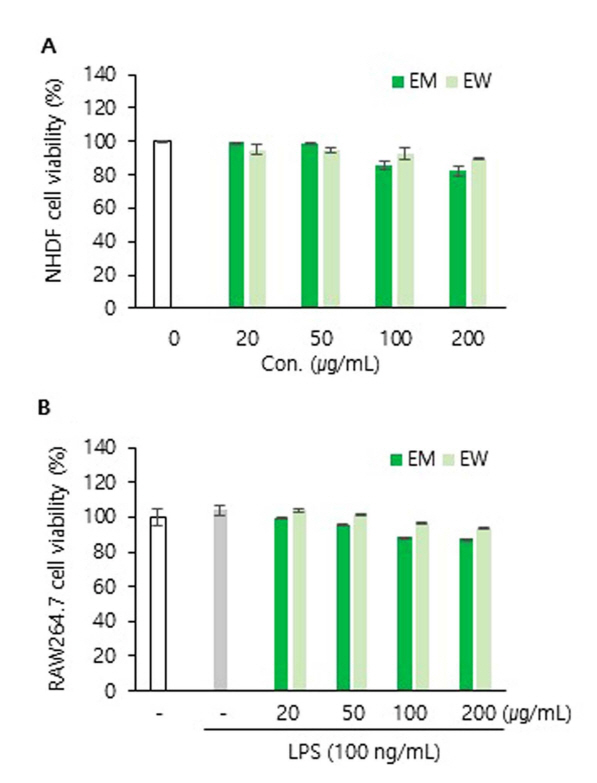
2. Cell viability

인간의 섬유아세포와 쥐의 면역세포를 대상으로 천근초 추출물 EM과 EW의 세포 생존율을 MTT assay를 수행한 결과 Fig. 1과 같다. 먼저 섬유아세포만 배양한 대조군을 100%로 기준하였을 때 각 추출물 농도 20, 50, 100 그리고 200 μg/mL에서 EM 처리에 의한 생존율은 99.2%, 98.9%, 85.4%, 82.1%, EW는 95.1%, 94.6%, 92.4%, 90.0%로 나타났다. 또한 항염 실험을 위해 수행한 RAW264.7 세포의 경우 동일한 농도 조건하에서 EM은 99.5%, 95.7%, 88.2%, 87.5%, EW는 103.8%, 101.1%, 96.7%, 93.4%의 생존율을 보였다. 천근초 에탄올추출물은 LPS에 의한 BV2 microglia의 세포보호 효능(Ding et al., 2016)과 rat 구강점막 자극(acute oral toxicity study)에 의한 사망률, 발병률, 일반 행동변화에 영향을 미치지 않았다(Parmar et al., 2019). 따라서 본 연구에서는 EM과 EW 모두 세포독성 실험과 동일한 최저 20 μg/mL에서부터 최고 200 μg/mL의 농도 범위를 설정하여 모든 실험을 진행하였다.

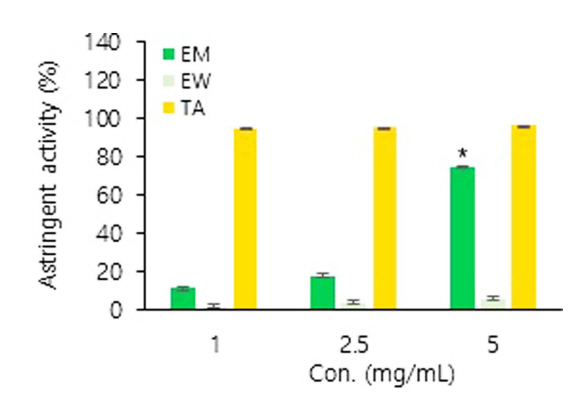
3. Astringent activity

피부에서 수렴 작용은 피부와 점막 혈관 수축, 점액 분비 억제, 모공 수축을 통해 팽팽한 피부 결을 나타내는 효과가 있다 . 천근초 아세톤추출물의 성분분석 결과 가수 분해형 탄닌(hydrolyzable tannin) isomallotinic acid 성분을 포함한 총 16개의 탄닌 화합물이 포함되어 있다고 알려져 있어(Lee et al., 1990), EM과 EW의 수렴활성을 확인한 결과 Fig. 2와 같다.
대표적인 수렴제인 탄닌산(Tannic acid, TA)은 1 mg/mL의 저농도에서 94.77%의 수렴활성을 보였다. 천근초 두 추출물의 수렴 활성은 모두 추출물 농도 의존적으로 수렴활성이 증가하였으며, 5 mg/mL 농도 조건에서 EM의 활성이 EW보다 12배 높게 나타났다. 특히 최고 농도 5 mg/mL일 때, EM의 활성이 75.03%로 양성대조군인 탄닌산 96.54%와 유사함을 확인 하였다.

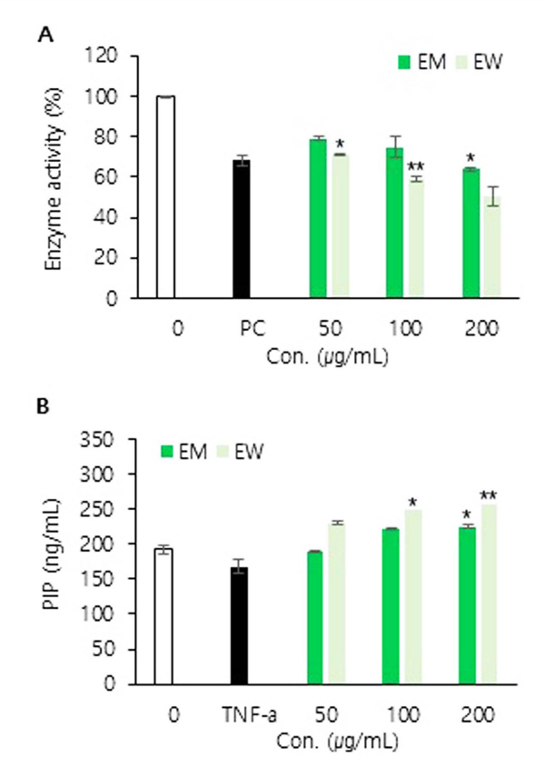
4. Anti-elastase activity

Elastase는 진피 내 피부 탄력을 유지하는 기질 단백질인 elastin을 분해하는 백혈구 과립효소 중 하나로 피부세포 기저층의 망상 조직을 파괴시켜 피부의 주름 및 탄력성 소실 등을 유발한다(Takeuchi et al., 2010). 천근초 추출물의 elastase 효소 저해 수준을 효소면역측정법(EIA, Enzyme immunoassay)으로 수행한 결과 Fig. 3A와 같다. 양성대조물질로 사용된 protease inhibitor인 elastatinal을 사용한 결과 31.7%로 효소 활성이 억제되었다. EM과 EW 추출물의 최고농도 200 μg/mL에서 각각 36.2%, 49.3%로 효소 활성이 감소하였으며 이는 elastatinal이 단일물질임을 감안할 때 천근초 추출물의 elastase 효소 활성 저해능이 매우 우수함을 나타낸다.

5. Procollagen synthesis ability

진피의 결합조직 내에 존재하는 collagen은 세포 내에서 procollagen이라는 전구물질로 합성된 후 세포 외로 분비되어 collagen 섬유로 중합되는데, procollagen의 N-말단 및 C-말단의 propeptide가 endopeptidase에 의해 유리되는 것으로 밝혀졌다(Seo et al., 2017). 따라서 procollagen C 말단 propeptide의 함량을 측정한 결과 Fig. 3B와 같다. 세포만 배양한 무처리군의 PIP 생성량이 192.55 ng/mL 일 때, 양성대조군 TNF-α 처리 시 167.79 ng/mL로 감소하였다. 모든 추출물 농도 의존적으로 PIP 생성이 증가하였으며 200 μg/mL 농도에서 무처리군과 비교하여 EW 추출물에 의한 PIP 생성이 EM보다 1.3배 높았다.

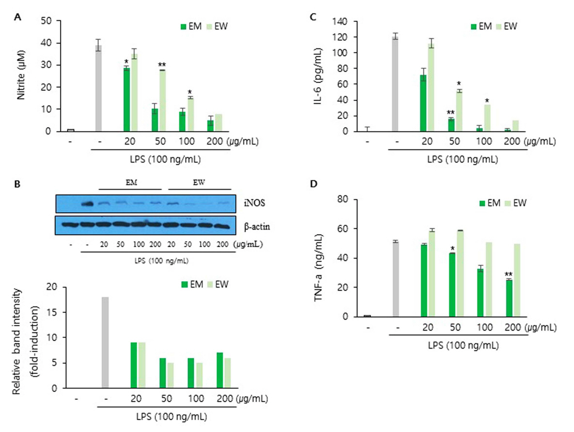
6. Anti-inflammatory activity

NO 생성을 조사하기 위해 아질산염(nitrite, NO2 -)을 주로 측정하는데 이는 NO의 비휘발성 분해 산물이며 매우 안정한 물질로 Griess 시약과 diazotization 반응을 통해 시료를 분홍색으로 변색 시킨다. 따라서 EM 또는 EW 추출물에 의한 세포 배양액 내 아질산염 검출을 측정한 결과 Fig. 4와 같다. 염증유도 물질 LPS에 의해 증가된 NO 양은 세포만 배양한 대조군에 비해 약 40배 증가하였으며, 이러한 조건에서 두 가지 추출물 처리에 의한 NO 생성 감소는 EM에 의해 더욱 뚜렷하게 나타남을 확인하였다. 천근초와 동일한 속(genus) Euphorbia hirta 추출물의 항염 기전 연구 결과에서 NO 합성 효소인 iNOS(inducible nitric oxide synthase)의 단백 발현 감소를 통한 NO 생성량 억제기전 결과(Shih et al., 2010)와 동일하게 본 연구에서 확인된 NO 생성 저하는 iNOS 효소 감소에 의한 것임을 확인하였다.
또한 염증성 사이토카인 TNF-α, IL-6의 분비량을 ELISA analysis로 수행한 결과 Fig. 4와 같다. 그 결과 LPS 자극에 의해 급격히 증가된 사이토카인 TNF-α, IL-6의 분비가 두 추출물 농도 의존적으로 저하됨을 확인하였다. 이러한 결과는 Carrageenan induced paw edema 동물 모델에서 천근초 에탄올추출물에 의한 항염 연구결과(Garipelli et al., 2012)와 맥을 같이 하며, 본 연구 결과가 세포 수준에서 천근초의 항염기전에 대한 분자생물학적 기초자료로서 가치가 높다고 판단된다.

IV. 결 론

본 연구에서는 천근초 80% 메탄올추출물(EM)과 열수추출물(EW)의 항염, elastae 효소 활성 저해능, 콜라겐 생성 및 수렴 활성 효능을 평가하여 항노화 화장품 원료로서의 가능성을 조사하였다. 천근초 건조 시료를 80% 메탄올과 60oC 열수 두 가지 용매로 추출한 결과 최종 수율은 EM 18.548%, EW 20.161%로 확인되었다. 수렴활성 결과 EM의 활성이 EW보다 높았으며 고농도(5 mg/mL) 조건에서 EM의 수렴활성은 양성대조군인 탄닌산과 유사하였다. Elastase 효소 활성 저해능은 추출물 최고농도 200 μg/mL에서 36.2%, 49.3%로 EM보다 EW가 높았으며 EW의 효소 저해수준은 양성대조물질인 elastatinal(31.7%)보다 더 우수하였다. 콜라겐 생성을 PIP로 측정한 결과 두 가지 추출물 농도 의존적으로 증가하였으며 무처리군과 비교하였을 때 EW가 EM보다 1.3배 높게 확인되었다. 천근초 추출물의 항염 효능을 조사하고자 쥐 대식세포(RAW264.7)에 염증 유도물질 LPS로 자극시킨 염증모델을 이용하여 조사한 결과 LPS에 의해 유도된 NO 생성이 EM과 EW 처리 후 감소하였다. 특히 EM 농도에 따른 NO 생성이 급격하게 감소하였는데 이는 세포 내 NO 생성에 필요한 iNOS 효소의 단백질 수준을 immunoblotting assay로 분석한 결과 각 추출물이 iNOS의 발현을 억제하여 NO 생성을 저해하는 것으로 확인되었다. 또한 대표적인 염증성 싸이토카인 TNF-α와 IL-6의 분비량이 천근초 추출물에 의해 감소하였다. 따라서 천근초 추출은 피부 염증과 노화를 억제시키는 항노화 화장품 원료로서의 활용가치가 높을 것으로 판단된다.

Fig. 1.
Cell viability of E. thymifolia extracts on (A) NHDF and (B) RAW264.7 cells. The cytotoxicity of extracts in cells was analyzed by MTT assay in triplicate and expressed in percentage of control.
JKSC-2022-28-3-525f1.jpg
Fig. 2.
Astringent activity of E. thymifolia extracts. Tannic acid (TA) was used as the positive control.
Data are represented as the mean±SD (n=3). Statistical differences are presented *p<0.05
JKSC-2022-28-3-525f2.jpg
Fig. 3.
Anti-aging effect of E. thymifolia extracts. (A) Elastase inhibitory activity with elastatinal for positive control (PC) inhibitor, (B) procollagen type I production (PIP). Data are represented as the mean±SD (n=3). Statistical differences are presented *p<0.05, **p<0.01
JKSC-2022-28-3-525f3.jpg
Fig. 4.
Effects of E. thymifolia extracts on LPS-stimulated inflammatory response in RAW264.7 cells.
(A) The medium was collected to determine the nitrite level using Griess reagent, (B) iNOS expression was detected by immunoblotting. (C and D) The TNF-α and IL-6 levels were detected using ELISA kits. Data are represented as the mean±SD (n=3). Statistical differences are presented *p<0.05, **p<0.01
JKSC-2022-28-3-525f4.jpg

References

Ding, H. Y., Wu, P. S., & Wu, M. J. (2016 Cleome rutidosperma and Euphorbia thymifolia suppress inflammatory response via upregulation of phase II enzymes and modulation of NF-B and JNK activation in LPS-stimulated BV2 microglia. International Journal of Molecular Sciences, 17(9), 1420-1435. doi:10.3390/ijms17091420.
crossref pmc
Durai, Muthumani G., Vijay, Anand A., & Manikandan, R. (2013). Antioxidant, antihelmintic and antimicrobial activity of Euphorbia thymifolia Linn whole plant. International Journal of Current Microbiology and Applied Sciences, 2(1), 66-79.
Enzo Life Sciences: Neutrophil Elastase Colorimetric Drug Discovery Kit. Available from: http://www.enzolifesciences.com/BMLAK497/neutrophil-elastasecolorimetric-drugdiscovery-kit. [Last accessed on 2020 Feb 22].
Ernst, M., Grace, O. M., Saslis-Lagoudakis, C. H., Nilsson, N., Simonsen, H. T., & Ronsted, N. (2015). Global medicinal uses of Euphorbia L. (Euphorbiaceae). Journal of Ethnopharmacology, 176:90-101.
crossref pmid
Garipelli, N., Runja, C., Poturi, N., & Pigili, R. (2012). Anti-inflammatory and anti-oxidant activities of ethanolic extract of Euphorbia thymifolia Linn whole plant. International Journal of Pharmacy and Pharmaceutical Sciences, 4(3), 516-519.
Gurava, Rdeey K., Anuradha, V., & Samba, Siva Rao V. (2017). Phytochemical and antioxidant activities of methanolic extact of the plant Euphorbia thymifolia Linn. Natural Products Chemistry & Research, 5(8), 1-3.
Ji, S. J., & Oh, B. U. (2009). Taxonomic reviews on three taxa of Korean Euphoria L. Korean Journal of Plant Taxonoy, 39(2), 100-106.
Kim, H. S., & Ko, K. S. (2021). The effect of ginseng flower extract on the skin aging inhibition. Journal of The Korean Society of Cosmetology, 27(3), 688-694.
Kim, J. A., Choi, J. Y., Son, A. R., Park, S. H., Xu, G. H., Lee, J. G., Oh, I. S., Kim, J. J., Chang, H. W., Chung, S. R., Jang, T. S., & Lee, S. H. (2004). Inhibitory effect of some natural polyphenols isolated from Euphorbiaceae plants on melanogenesis. Korean Journal of Pharmacognosy, 35(2), 157-163.
Kim, J. S., & Lee, J. A. (2021). A study on anti-oxidant and anti-wrinkle effect of supercritical fluid extraction of black carrot as a functional cosmetic material. Journal of Convergence for Information Technology, 11(12), 236-243.
Kim, J. Y., Lee, J. A., Yoon, W. J., Oh, D. H., Jung, Y. H., Lee, W. J., & Park, S. Y. (2006 Antioxidative and antimicrobial activities of Euphorbia jolkini extracts. Korean Journal of Food Science and Technology, 38(5), 699-706. http://www.koreascience.or.kr/article/JAKO200603042166775.j.
Kim, N. S., Kim, K. C., & Oh, W. Y. (2019). Study of effect of Discorea japonica Thunb ethanol extract on anti-inflammation and anti-wrinkle. Journal of The Korean Society of Cosmetology, 25(4), 947-954.
Lee, S. H., Kanaka, T., Nonaka, G. I., & Nishioka, I. (1990 Hydrolysable tannins from Euphorbia thymifolia. Phytochemistry, 29(11), 3621-3625. doi:10.1016/0031-9422(90)85288-Q.
Li, C., & Wang, M. H. (2014 In vitro biological evaluation of 100 selected methanol extracts from the traditional medicinal plants of asia. Nutrition Research and Practice, 8(2), 1551-157. doi: 10.4162/nrp.2014.8.2.151.
Mali, P. Y., & Panchal, S. S. (2020 A review on phyto-pharmacological potentials of Euphorbia thymifolia L. Ancient Science of Life, 32(3), 165-172. doi:10.4103/0257-7941.123001.
Manyou, Y., Irene, G., Joao, R., & Ana, B. (2021 Phytochemical and antioxidant analysis of medicinal and food plants towards bioactive food and pharmaceutical resources. Scientific Reports, 11(1), 10041-10054. doi:10.1038/s41598-021-89437-4.
pmid pmc
Muthumani, D., Hedina, A., Kausar, J., & Anand, V. (2016 Phytopharmacological activities of Euphorbia thymifolia Linn. Systematic Reviews in Pharmacy, 7(1), 30-34. doi:10.5530/srp.2016.7.4.
Parmar, G., Pundarikakshudu, K., & Balaraman, R. (2019 Anti-anaphylactic and antiasthmatic activity of Euphorbia thymifolia L. on experimental animals. Journal of Traditional and Complementary Medicine, 9(1), 60-65. doi:10.1016/j.jtcme.2018.03.002.
crossref pmid
Seo, W. Y., Kim, J. H., Baek, D. S., Kim, S. J., Kang, S., Yang, W. S., Song, J. A., Lee, M. S., Kim, S., & Kim, Y. S. (2017 Production of recombinant human procollagen type I C-terminal propeptide and establishment of a sandwich ELISA for quantification. Scientific Reports, 7(1), 1-13. doi:10.1038/s41598-017-16290-9.
crossref pmid pmc
Shih, M-F., Chen, Y-D., Shen, C-R., & Cherng, J. Y. (2010 A molecular pharmacology study into the anti-inflammatory actions of Euphorbia hirta L. on the LPS-induced RAW264.7 cells through selective iNOS protein inhibition. Journal of Nature Medicine, 64(3), 330-335. doi:10.1007/s11418-010-0417-6.
crossref
Takeuchi, H., Gomi, T., Shishido, M., Watanabe, H., & Suenobu, N. (2010 Neutrophil elastase contributes to extracellular matrix damage induced by chronic low-dose UV irradiation in a hairless mouse photoaging model. Journal of Dermatological Science, 60(3), 151-158. doi:10.1016/j.jdermsci.2010.09.001.
crossref pmid
Wang, Y. R., & Lee, J. A. (2018). The study of antioxidant and antiinflammatory effects of notoginseng root (NGR) hot water extracts. Journal of The Korean Society of Cosmetology, 24(5), 1014-1020.
Wunsch, E., & Heidrich, H. G. (1963 Zur quantitativen bestimmung der kollagenase. Hoppe-Seyler's Zeitschrift Für Physiologische Chemie, 333(Jahresband), 149-151. doi:10.1515/bchm2.1963.333.1.149.
crossref
Yoon, D. H., Han, C. W., Fang, Y. Y., Gundeti, S., Han, I. S., Song, W. O., Hwang, K. C., Kim, T. W., Sung, G. H., & Park, H. (2017 Inhibitory activity of Cordyceps bassiana extract on LPS-induced inflammation in RAW 264.7 cells by suppressing NF-κB activation. Natural Product Sciences, 23(3), 162-168. doi:10.20307/nps.2017.23.3.162.
crossref
TOOLS
METRICS Graph View
  • 2 Crossref
  •  0 Scopus
  • 1,311 View
  • 13 Download
Related articles


ABOUT
BROWSE ARTICLES
EDITORIAL POLICY
FOR CONTRIBUTORS
Editorial Office
69 Gwangju Yeodae-gil, Gwangsan-gu, Gwangju, 62396, Korea
Tel: +82-10-2825-6735   Fex: +82-62-950-3797    E-mail: beauty2007@hanmail.net                

Copyright © 2024 by Korean Society of Cosmetology.

Developed in M2PI

Close layer
prev next