J Korean Soc Cosmetol > Volume 30(2); 2024 > Article
괄루근 추출물이 콜라겐 생성에 미치는 영향

Abstract

This study was conducted to study the effects of Trichosanthis Radix water extract (TR-W) and Trichosanthis Radix 60% ethanol extract (TR-E) on collagen production. As a result of conducting a cytotoxicity experiment on HDFn cells and RAW 264.7 cells, the experiment was conducted at a concentration of TR-W and TR-E at 100 mg/ml, which does not significantly affect the survival rate in HDF and RAW 264.7 cells. The effect on collagen synthesis ability showed an increase of 52% for the positive control group L-AA (vitamin C) 52% for TR-W 25% and TR-E 36% at 100 mg/ml proving its effect on collagen synthesis ability. In terms of inhibition of MMP-1 expression related to collagen damage L-AA (vitamin C) a positive control group, showed an inhibitory effect of 54%, and TR-W and TR-E showed an inhibitory effect of 72% and 68% at 100 mg/ml, thereby inhibiting collagen decomposition enzyme. MMP-1 was stably inhibited. Compared to Control, the free radical scavenging activity of L-AA (vitamin C) a positive control group, showed a scavenging activity of 108.4%, and in TR-W and TR-E, it showed an inhibitory effect of 93.4% and 98.2% at 100 mg/ml. In NO related to inflammation, showed an anti-inflammatory effect of 67.4% and 50.2% at a concentration of 100 mg/ml of TR-W and TR-E compared to the control group, which is expected to have a positive effect on collagen production due to inflammation. This study The results are judged to have the highest utility value as a functional cosmetic material considering the stability of the skin in the future.

I. 서 론

주름은 피부노화 정도의 피부상태를 알려주는 기준으로 피부 노화의 원인은 주로 유전적 원인인 내인성 노화와 생활환경 등 외부환경에 의한 외인성 원인으로 나뉜다(Kim et al., 2007). 내인성 노화는 자외선(UV) 등 외부 자극에 의해 피부에서 주로 나타나며 피부의 각질층과 진피층에 도달하여 Collagen, elastin, fibronectin, proteoglycans 등에 영향을 주어 주름생성에 영향을 미친다(Fisher et al., 1996).
UVB는 콜라겐 분해효소인 matrix metalloproteinase(MMP)의 과발현으로 콜라겐 생성 억제, 노화, 주름, 거친 피부, 건조 피부, 상처치유 지연 등이 피부 광노화의 주요 원인으로 알려져 있다(Kang et al., 2003).
광노화는 대표적인 주름 생성의 원인으로 콜라겐 생성 능력 저하와 콜라겐 분해의 촉진으로 알려져 있다. 콜라겐은 피부의 주요 구성성분으로 진피의 70~80%의 비중을 차지하고 있다(Yang et al., 2007).
콜라겐의 총량은 콜라겐의 합성과 분해에 의해서 정해진다. 콜라겐의 생산(collagen production)은 복잡한 과정을 거쳐 생산된다(Sorushanova et al., 2019).
진피세포에서는 프리콜라겐(precollagen)과 프로콜라겐(procollagen) 등이 콜라겐을 만들어 세포밖으로 분비하고 교차연결(cross link)에 의해 안정화를 이루며 탄력성을 부여하는 구조체를 이룬다. 이러한 지지력으로 피부의 주름 억제와 탄력을 유지하는데 중요한 요소로 작용을 한다(Mouw et al., 2014). 또한, 콜라겐 단백질은 피부의 상처회복, 세포이동, 기관형성 및 여러 염증작용에도 관여한다고 알려져 있다(Her et al., 2019).
노화가 진행되면 피부 각질층은 피부장벽 기능이 약해지게 되어 수분 보유 능력 감소와 외부유해물질에 의한 피부 민감도가 증가하여 피부트러블이 증가한다(Netzel et al., 2002). 진피층에서는 콜라겐 및 fibronectin과 같은 extracellular matrix(ECM)의 기능이 저하되면서 피부는 얇고, 탄력이 감소하면서 피부가 위축되면서 주름이 발생한다(Panwar et al., 2018).
주름발생의 원인으로 알려진 MMP-1은 주로 진피층에서 있는 콜라겐의 분해를 촉진하여 피부 탄력을 감소시켜 주름을 발생시킨다. 이러한 이유로, MMP-1의 억제는 주름생성억제에 중요한 요소가 된다(Qin et al., 2017: Freitas et al., 2017).
하늘타리(Trichosanthes kirilowii Maxim)는 박과(Cucurbitaceae) 하늘타리속(Tricho -santhes)에 속하는 다년생덩굴 식물로 우리나라 중부이남에서 자생하고 있다(Lee, 2000).
한국, 일본, 몽골, 대만 등에서 자생하는 박과의 다년생 덩굴성 식물로 잎은 둥근모양이며, 단풍잎처럼 5~7갈래로 갈라지며 잎면에 톱니가 보이고, 아랫부분은 심장형이며 잎표면에 짧은 털이 있다(Jung & Shin, 1998; Huang et al., 2000).
하늘타리 뿌리에서는 녹말성분이 많아 식용하였으며, 열매, 씨앗 및 뿌리등을 약재로 사용하고 있다. 한방에서는 열매와 뿌리를 해열, 해수, 이뇨, 변비, 어혈, 황달, 피부병, 결핵, 유방암, 적백리, 당뇨병등의 치료에 사용한다(Zhu, 1998).
이중 한약재로 사용되는 괄루근(Trichosanthis Radix)은 하늘타리 또는 쌍변괄루(Trichosanthes rosthornii Harms)라 불리우며, 뿌리의 표피를 제거한 것을 말한다.
성분은 trichosanthin(TCN), TAP-29, trichok irin, α-kirilowin, β-kirilowin,karasurin과 같은 다양한 Ribosome-inactivating protein (RIP)이 함유되어 있고, 이러한 물질은 염증을 억제하고, 산소활성를 제거하여 염증 반응을 억제하는 것으로 알려져 있다(Byers et al., 1990).
이에 본 연구자는 괄루근의 물 추출물과 70% 주정 추출물을 사용하여 자외선 조사로 자극하여 MMP-1발현 변화, 항염 작용, 항산화능과 콜라겐 합성에 미치는 영향을 알아보고자 한다.

II. 재료 및 방법

1. 약재

실험에 사용된 괄루근(Tricosanthis Radix: TR)은 한국 옴니허브에서 구입하였다. 약재는 초음파 세척기(Branson, U.S.A.)를 이용하여 불순물을 제거하여 실험에 사용하였다. 건조중량 1.2 kg를 냉각수 환류하여 4시간씩 3회 추출하고, 여과, 감압 농축하여 105.26g으로 8.77% 수득율을 얻었다(TR-W). 건조중 1.2 kg를 60% 에탄올에서 4시간씩 환류하여 3회 추출하여 여과, 감압 농축하여 60% 주정 추출물 99.71 g으로 8.31%의 수득율을 나타내었다(TR-E).

2. 세포 배양조건

섬유아세포(human dermal fibroblast neonatal: HDFn, ACTT)와 대식세포(RAW 264.7 : macrophage. Korean Cell Line Bank, Korea)를 사용하였다. 세포주는 DMEM 배지에 10%의 FBS를 첨가하여 배양하였고, 배양된 세포는 PBS을 이용하여 3회 세척하였다. Trypsin-EDTA를 투여하여 배양시킨 후, 현미경을 이용하여 세포의 분리를 확인하고 10% FBS가 첨가된 배지로 trypsin-EDTA를 희석시켰다. 세포를 동량의 trypan blue와 혼합한 후 Hemocytometer를 이용하여 현미경으로 세포 수를 측정하였다.

3. 시약 및 기기

1) 시약

본 실험에 사용된 시약은 Lipopolys accharide(LPS), Dulbecco's modified Eagle's medium(DMEM), fetal bovine serum, penicillin, phosphate buffered saline solution(PBS), primary antibody(anti-MMP-1 mouse antibody), Griess reagent (Sigma-Aldrich, U.S.A.), MTT(3-[4,5-dimethylthiazol-2-yl]-2,5-diphenyl tetrazolium bromide)로 부터 구입하여 사용하였고, 이외의 시약은 특급시약을 사용하였다. 사용된 기기는 Centrifuge(Hanil, Korea), vortex mixer(Vision Scientific Co., Korea), CO2 incubator(Sanyo, Osaka, Japan), rotary vacuum evaporator(Eyela, Japan), deep freezer(Ilshin Lab Co., Korea), microplate reader(Bio-Rad, U.S.A.), ELISA reader(Spectrostarnano, BMG Labtech, Offenburg, Germany), water bath (In-Tron biotech, Korea), ice-maker(Vision Scientific Co., Korea), clean bench (Jeiothec, Korea), ultra soniccleaner(Branson, U.S.A.), Bio-Plex200(Bio-Rad, U.S.A.), spectrofluorometer(Dynex, U.K.) 등이다.

4. 실험방법

1) 세포 생존율 측정

HDFn 세포와 RAW 264.7 세포는 한국세포주은행(KCLB, Seoul, Korea)에서 분양받아 DMEM 배지: 10% FBS를 사용하여 37℃, 5% CO2 incubator에서 배양하였다. 시료의 세포독성은 MTT(Kim, 2003) assay를 사용하여 실험하였다. 96 well plate에 세포를 1×10⁴ cells/well씩 접종하고 시료를 200 ㎕씩 가한 후, 48시간 동안 배양하였다. 이 후, MTT 용액(3 mg/ml)을 50 ㎕씩 첨가하고 CO2 incubator에 넣어 4시간 동안 반응시켰다. 상등액을 제거하고 dimethylsulfoxide(DMSO) 150 ㎕씩 첨가하고 595 nm(Sunrise, Tecan, Switzerland)에서 흡광도를 측정하였다.

2) 콜라겐 합성능 측정

HDFn를 5×10⁴ cells/well로 24-well plate에 접종한 후 DMEM:10% FBS에서 37℃ incubator에서 24시간 동안 배양하여 사용하였다. Trypsin-EDTA를 투여하여, 세포를 분리시키고, 일정양의 세포액과 동량의 trypan blue와 혼합하였다. 혼합된 세포를 hemocytometer를 이용하여 세포수를 측정였다. 시료는 0.02, 0.05%로 serum이 없는 DMEM 배지에서 24시간 배양 후 콜라겐 생성 정도를 PIP assay kit(Takara)를 이용하여 측정하였다. 이때 양성대조군으로 L-AA(vitamin C)를 처리하여 콜라겐 합성 정도를 평가하였다(Parfitt, 1987).
검액은 배양액을 PBS에 희석하여 사용하며 콜라겐 정량 시험법으로 진행하였다. 프로콜라겐-I, C-펩타이드(PIP) EIA kit(Takara Bio Inc.)에 antibody-POD conjugate solution을 넣고, 검액 및 표준시료(Standard : 0, 40, 80, 160, 320, 640 ng/㎖)를 각각 20 ㎕씩 첨가하여 Mix후 밀봉하여 37℃에서 3시간 반응 후 1N H2SO4를 넣어 반응을 중지시키고 450 nm에서 흡광도를 측정하였다.

3) 콜라겐 분해 효소 발현 억제 기전 평가

HDFn 5×10⁴ cells/well에 농도별로 접종한 후 DMEM:10% FBS조건에서 37℃ incubator에서 24시간 동안 배양하였다. UVA를 6 J/cm2로 조사하고 FBS가 첨가되지 않은 배지로 교환 후 시료를 처리하고 24시간동안 배양하였다. 24시간 후에 상층액으로 BIOTRAK ELISA human MMP-1 nRTN2601을 이용하여 450 nm에서 흡광도를 측정하였다.

4) DPPH 자유 라디칼 소거 활성

자유 라디칼 소거 활성은 Ikeda 등의 방법에 준하여 측정하였다. 각 시료를 일정 농도가 되도록 에탄올에 넣어 200 ㎕로 맞추고, 에탄올에 녹인 0.1 mM 2,2-diphenyl-1-(2,4,6-trinitrophenyl)hydrazin-1-yl(DPPH) solution을 200 ㎕씩 가한 후, 교반하고 실온에서 30분 동안 방치한 다음 519 nm(Sunrise, Tecan, Switzerland)에서 흡광도를 측정하였다. 양성 대조군은 α-토코페롤을 사용하여 소거활성능을 대조군과 비교하여 감소율로 나타내었다.

5) Nitric oxide(NO) 생성 억제능

항염증 효과를 알아보기 위하여 NO 생성 억제능을 측정하였다. RAW 264.7 세포를 96well plate에 5×10⁴ cells/well의 농도로 분주하고, 24시간 동안 배양 후 lipopolysaccharide(LPS) 1 ㎍/㎖ 농도로 처리된 배지에 TR-W, TR-E를 농도별로 처리하여 48시간 동안 배양하였다. 96well plate에 Griess reagent(Sigma-Aldrich, U.S.A.)와 배양된 세포 배양 상층액을 가하여 10분 반응시킨 후 microplate reader(Synergy HT; BioTek Instruments, U.S.A.)를 이용하여 540 nm에서 흡광도를 측정하였다.

5. 통계

실험으로부터 얻은 결과는 mean ± standard error로 기록하였다. 유의성 검증은 Student's T-test 분석법을 이용하여 작성하였다. 유의성 검증은 Student’s t-test를 실시하였고, p값이 0.05 미만일 때 통계적으로 유의한 차이가 있는 것으로 판정하였다.

III. 결과 및 고찰

1. 세포독성

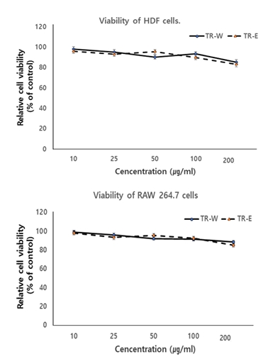
HDF와 RAW 264.7 세포에 대한 TR-W와 TR-E의 영향을 MTT assay를 이용하여 독성을 확인하였다. HDF와 RAW 264.7 세포에 TR-W와 TR-E를 각각 10, 25, 50, 100, 200 ㎍/㎖의 농도별로 처리하고, 시료를 처리하지 않은 세포를 대조군으로 48시간 동안 배양하여 비교하였다. 그 결과 HDF와 RAW 264.7 세포에서 TR-W와 TR-E에서 100 ㎍/㎖ 이하에서 10% 이하의 독성을 나타내어 세포에 거의 영향을 주지 않는 것으로 확인하여 TR-W와 TR-E의 세포의 생존율에 크게 영향을 미치지 않는 100 ㎍/㎖ 이하로 실험을 진행하였다(Fig. 1).

2. 콜라겐 합성능

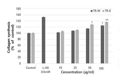
피부는 표피, 진피, 피하조직으로 나뉘어 있으며, 이중 진피는 섬유성분, 기질성분으로 구성되어 있고 섬유성분으로 존재하는 콜라겐은 진피의 90%를 차지하고 있다(Bradford, 1976). 콜라겐은 피부의 섬유아세포에서 발현되는 단백질로 피부, 뼈 및 치아등 우리 인체의 유기 물질의 대부분을 구성하고 있으며 피부의 견고성, 결합조직의 저항력, 조직의 결합력, 세포 증식, 세포의 지탱 등 다양한 기능을 가지고 있다(Kim & Lee, 2015). 콜라겐은 피부에서 주름 형성과 탄력에 밀접한 연관이 있고 콜라겐이 부족할 경우 탄력이 감하면서 주름을 유발할 수 있다(Kim & Lee, 2015).
HDF 세포에 대한 TR-W와 TR-E의 콜라겐 생성 합성능을 측정하기 위하여 HDF 세포에 48시간 동안 처리한 후 배양 상층액을 취하여 콜라겐의 함량을 ELISA로 측정한 결과, Control에 비하여 양성대조군인 L-AA(vitamin C)는 152%의 증가를 나타내었고, TR-W와 TR-E에서 100 ㎍/㎖에서 25%, 36%의 증가를 나타내었으며 TR-E가 TR-W에 비하여 유의적인 증가를 나타내어 효과를 입증하였다. 이와 같은 결과는 콜라겐 생성 촉진 효과를 확인한 것으로 이를 첨가한 화장품 개발 등의 기능성 화장품 소재로서의 적용 가능성이 있다고 사료된다(Fig. 2).

3. MMP-1 발현억제능 측정

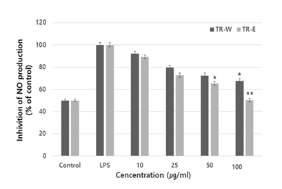
콜라겐은 결합조직의 탄력을 유지에 필수적 성분으로 UV나 외부자극에 의해서 콜라겐은 분해되고 재합성이 되지 않으면 피부조직에 손상을 가져오게 만드는 역할을 한다(Tsuji et al., 2001). 콜라겐을 분해하는 효소는 다양하지만 그중 MMP-1 (matrix metalloproteinase 1)이 가장 대표적이다(Choi, 2007).
MMP-1은 주로 진피에서 콜라겐을 분해하는 효소로 알려져 있으며 과도하게 발현되면 콜라겐의 분해를 촉진하여 피부에서 교원질의 결핍을 초래하여 피부 주름, 탄력 상실 및 수분 소실 등 진피세포의 노화반응 촉진에 작용한다(Kang et al., 2003). 이러한 이유로 MMP-1 효소의 활성 저해는 주름생성 억제에 중요한 역할을 할 수 있으며, 콜라겐 보호가 피부 조직의 탄력을 유지하고 지속시켜 주름생성의 진행을 억제할 수 있다고 알려져 있다(Jung et al., 2014).
UVA에 의해 발현이 증가되는 MMP-1에 TR-W 와 TR-E이 미치는 영향을 알아보고자 섬유아세포(HDF-n)에 6.3 J/cm2 UVA를 조사하여 24시간 동안 배양한 후 MMP-1 발현 저해 효과를 측정한 결과, Control에 비하여 양성대조군인 L-AA(vitamin C)는 54%의 저해효과를 나타내었고, TR-W와 TR-E에서 100 ㎍/㎖에서 72%, 68%의 저해효과를 나타내었으며 TR-E에서 유의적인 효과를 나타내었다(Fig. 3). MMP-1은 UV로 인한 피부 손상과 조기 노화를 일으키는 중요한 역할을 하므로 TR-W와 TR-E에 의한 MMP-1 억제는 광노화 억제에 중요한 인자로 작용할 것으로 사료된다(He et al., 2021).

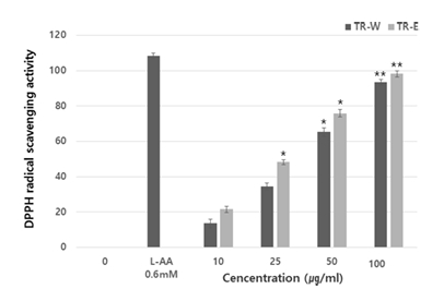
4. DPPH 자유 라디칼 소거 활성

전자공여능 측정에 사용된 1,1-diphenyl-2-picrylhydrazyl은 매우 안정한 자유 라디칼로 517 nm에서 특징적으로 광흡수를 나타내는 보라색 화합물이다(Aoshima et al., 2004). 자유라디칼은 하나의 전자를 가지고 있는 불안전한 분자로 인체 내 지질 또는 단백질 등과 결합하여 노화를 일으키기가 용의하며, 페놀성 화합물의 경우 자유라디칼을 환원시키거나 상쇄시키는 능력으로 인체 내에서 자유 라디칼에 의한 항노화 작용을 한다. 이를 항산화제라 한다(Marklund & Marklund, 1974). DPPH는 알코올 등의 유기용매에 안정된 상태를 유지하고 있다(Ikeda & Fukuzumi, 1977) 항산화 기전 중 proton-radical scavenger에 의하여 탈색되기 때문에 항산화 활성을 육안으로 쉽게 관찰할 수 있는 장점이 있어 다양한 천연소재로 항산화 물질을 검색하는데 많이 이용되고 있다(Song et al., 2016). TR-W와 TR-E를 다양한 농도에서 DPPH 라디칼 소거능을 측정한 결과, Control에 비하여 양성대조군인 L-AA(vitamin C)는 108.4%의 소거능을 나타내었고, TR-W 와 TR-E에서 100 ㎍/㎖에서 93.4%, 98.2%의 저해효과를 나타내었으며 TR-W는 50, 100 ㎍/㎖에서 TR-E는 25, 50, 100 ㎍/㎖에서 유의적인 소거능을 나타내었다(Fig. 4).

5. 항염

피부는 과도한 자외선 노출시 산화작용에 의해 피부에 산화적 스트레스를 축척하여 염증 반응을 통해 콜라겐 분해, DNA 변이등 피부손상을 야기한다(Inui et al., 2008). NO(nitric oxide)는 Macrophage가 분비하는 세포독성 물질로(Gabay, 2006), 과량 생성 시 조직과 신경 손상 유발, 유전자 변이 유도, 부종 유발 등 과도한 염증을 일으킨다(Im, 2014). NO는 체내에서 세균과 종양을 제거, 신경전달 조절, 혈압 조절등 다양한 역할을 담당하는 중요한 인자로 알려져 있다(Lee et al., 2014). 마우스의 대식세포인 RAW 264.7 세포를 LPS로 자극하여 항염증 효과를 확인한 결과, TR-W와 TR-E를 다양한 농도에서 NO 억제효과를 나타내었고, 100 ㎍/㎖에서 67.4%, 50.2%의 저해효과를 나타내었으며 TR-W는 100 ㎍/㎖에서 TR-E는 50, 100 ㎍/㎖에서 유의적인 억제효과를 나타내었다(Fig. 5).

IV. 결 론

본 연구는 TR-W과 TR-E이 콜라겐 생성에 미치는 영향에 대하여 연구를 진행하였다. 세포의 독성을 확인하기 위해 HDFn 세포와 RAW 264.7 세포독성 실험을 진행한 결과, HDF와 RAW 264.7 세포에서 TR-W와 TR-E가 생존율에 크게 영향을 미치지 않는 100 ㎍/㎖ 이하로 실험을 진행하였다.
콜라겐 합성능에 미치는 효과는 양성대조군인 L-AA(vitamin C)는 52%, TR-W, TR-E에서 100 ㎍/㎖에서 25%, 36%의 증가를 나타내어 콜라겐 합성능에 효과를 입증하였다. 콜라겐 손상과 관련된 MMP-1 발현 억제능에서는 양성대조군인 L-AA(vitamin C)는 54%, TR-W와 TR-E에서 100 ㎍/㎖에서 72%, 68%의 저해효과를 나타내어 콜라겐 분해효소인 MMP-1을 안정적으로 억제하였다. 자유라디컬 소거능도 Control에 비하여 양성대조군인 L-AA(vitamin C)는 108.4%의 소거능을 나타내었고, TR-W와 TR-E에서 100 ㎍/㎖에서 93.4%, 98.2%의 저해효과를 나타내었다. 염증과 관련된 NO에서는 대조군에 비하여 TR-W와 TR-E의 100 ㎍/㎖ 농도에서 67.4%, 50.2%의 항염효과를 나타내어 염증에 의한 콜라겐 생성에 긍적적인 효과를 줄것으로 사료된다. 이러한 연구결과는 향후 피부의 안정성을 고려하여 해당 기능성 화장품소재로써 활용가치가 가장 높을 것으로 판단된다.

Fig. 1.
Cytotoxicity of TR-W and TR-E on the HDF & RAW 264.7 cells. The results are presented as the mean ± S.D. of three independent experiments. Cell cytotoxicity was evaluated with the MTT assay. Mean values ± S.D. from triplicate separated experiments are shown. P values were calculated by ANOVA Test.
JKSC-2024-30-2-408f1.jpg
Fig. 2.
Collegen synthesis of TR-W and TR-E on the cell synthesis of human dermal fibroblasts. The cells were treated with various concentration of L-AA (vitamin C), TR-W and TR-E for 24h. The contents of Collagen type I in culture media were determined by the (PIP) EIA kit as detailed under the Materials and Methods. The results were expressed as the average of three intra assay with S.D. *p<0.05, **p<0.01 compared with control.
JKSC-2024-30-2-408f2.jpg
Fig. 3.
Inhibition effect of L-AA(vitamin C), TR-W and TR-E on MMP-1 activity. The cells were treated with various concentration of TR-W and TR-E for 24h. The results were expressed as the average of three intra assay with S.D. *p<0.05, **p<0.01 compared with control.
JKSC-2024-30-2-408f3.jpg
Fig. 4.
Free radical scavenging activity of L-AA (vitamin C), TR-W and TR-E on measured by DPPH assay. TR-W and TR-E were incubated with DPPH at 25℃ for 30 min and the absorbance at 519 nm was measured. The results were expressed as the average of three intra assay with S.D. *p<0.05, **p<0.01 compared with control.
JKSC-2024-30-2-408f4.jpg
Fig. 5.
Inhibition of NO production in RAW 264.7 cell treated with Paeonia lactiflora extract. The results were expressed as the average of three intra assay with S.D. *p<0.05, **p<0.01 compared with control.
JKSC-2024-30-2-408f5.jpg

References

Aoshima, H., Tsunoue, H., Koda, H., & Kiso, Y. (2004). Aging of whiskey increases 1,1-diphenyl-2-picrylhydrazyl radical scavenging activity. J. Agric. Food Chem, 52(16), 5240.
crossref
Bradford, M. M. (1976). A rapid and sensitive method for the quantitation of microgram quantities of protein utilizing the principle of protein-dye binding. Analytical Biochemistry, 72:248-254.
crossref pmid
Choi, Y. A. (2007). Signaling pathway of MMP-2 in fibroblast migration and chondrogenic differentiation, Department of Biology kyungpook University.
Fisher, G. J., Datta, S. C., Talwar, H. S., Wang, Z. Q., Varani, J., Kang, S., & Voorhees, J. J. (1996). Molecular basis of sun-induced premature skin ageing and retinoid antagonism. Nature, 379:335-339.
crossref pmid pdf
Freitas-Rodriguez, S., Folgueras, A. R., & Lopez-Otin, C. (2017). The role of matrix metalloproteinases in aging: Tissue remodeling and beyond. Biochimica et Biophysica Acta, Molecular Cell Research, 1864((11) Pt A), 2015-2025.
crossref pmid
Gabay, C. (2006). Interleukin-6 and chronic inflammation. Arthritis Research & Therapy, 8(2), 1917-1924.
He, Y. L., Xiao, Z., Yang, S., Zhou, C., Sun, S., Hong, P., & Qian, Z. J. (2021). A phlorotanin, 6,6'-bieckol from Ecklonia cava, against photoaging by inhibiting MMP-1, -3 and -9 expression on UVB-induced HaCaT keratinocytes. Photochem. Photobiol., 1131.
Her, Y., Shin, B. N., Lee, Y. L., Park, J. H., & Kim, D. W. (2019). Oenanthe Javanica Extract Protects Mouse Skin from UVB Radiation via Attenuating Collagen Disruption and Inflammation. International Journal of Molecular Sciences, 20(6), 1435.
crossref pmid pmc
Huang, Y., He, P., Bader, K. P., Radunz, A., & Schmid, G. H. (2000). Seeds of Trichosan the skirilowii, an energy-rich diet. Zeits chriftfür Natur for Schung C, 55(3-4), 189-94.
crossref
Ikeda, N., & Fukuzumi, K. (1977). Synergistic antioxidant effect of nucleic acids and tocopherols. J. Am. Oil Chem. Soc, 54(9), 360.
crossref pmid
Im, D. Y. (2014). Volatile compounds analysis of the extract from dried bark of Prunus sargentii and physiological activity of the main compound, benzaldehyde. Asian Journal of Beauty and Cosmetology, 12:155-162.
Inui, M., Ooe, M., Fujii, K., Matsunaka, H., Yoshida, M., & Ichihashi, M. (2008). Mechanisms of inhibitory effects of CoQ10 on UVB-induced wrinkle formation in vitro and in vivo. Biofactors, 32(1), 237.
crossref
Jung, B. S., & Shin, M. K. (1998). Encyclopedia of Traditional Drug, 2nd ed. Younglim Pub.; 960-3.
Jung, H. K., Jang, J. H., Ko, J. H., Kang, B. M., Yeo, J. H., Cho, J. H., Cho, H. W., Bean, C. G., Kim, S. C., & Jung, W. S. (2014). Whitening and anti-wrinkle effects of Korean native Dendrobium moniliforme methanol extract. Korean Journal of Medicinal Crop Sciences, 22:331-338.
crossref
Kang, S., Chung, J. H., Lee, J. H., Fisher, G. J., Wan, Y. S., Duell, E. A., & Voorhees, J. J. (2003). Topical N-acetyl cysteine and genistein prevent ultraviolet-light-induced signaling that leads to photoaging in human skin in vivo. J. Invest. Dermatol, 120:835-841.
crossref pmid
Kim, H. K., Kim, E. S., Choi, I. B., Kim, J., & Lee, S. J. (2003). The verification of the MTT assay on the viability of periodontal ligamental cells in rat molars through the histologic examination. J. Kor. Acad. Cons. Dent, 28(5), 385-391.
crossref
Kim, J. M., Jeon, S. W., Lee, W. G., Nam, H. J., & Kim, Y. B. (2010). Study of preventing methods for skin aging and wrinkles. Korean J. Oriental Physiology & Pathology, 24(4), 533.
Kim, N. Y., & Lee, H. Y. (2015). Effect of antioxidant and skin whitening of ethanol extracts from ultrasonic pretreated Abeliophyllum distichum Nakai. Korean Journal of Medicinal Crop Sciences, 23:155-160.
crossref
Kim, N. Y., & Lee, H. Y. (2015). Enhancement of anti-wrinkle activities of Abeliophyllum distichum Nakai through low temperature extraction process. Korean Journal of Medicinal Crop Sciences, 23:231-236.
crossref
Lee, H. J., Sim, B. Y., Bak, J. W., & Kim, D. H. (2014). Effect of Gami-sopungsan on infla- mmation and DNCB-induced dermatitis in NC/Nga in mice. Journal of Physiology & Pathology in Korean Medicine, 28:146-153.
crossref
Lee, Y. N.. (2000). New Korean strated plant book I Seoul, Kyohaksa, p.639-774.
Marklund, S., & Marklund, G. (1974). Involvement of the superoxide anion radical in the autoxidation of pyrogallol and a convenient assay for superoxide dismutase. Eur. J. Biochem, 47(3), 469.
crossref pmid
Mouw, J. K., Ou, G., & Weaver, V. M. (2014). Extracellular matrix assembly: a multiscale deconstruction. Nature Reviews. Molecular Cell Biology, 15(2), 771-785.
crossref pmid pmc pdf
Netzel, S., Mitola, D. J., Yanada, S. S., & Holmbeck, K. (2002). Collagen dissolution by keratinocytes requires cell surface plasminogen activation are matrix metalloproteinase activity. J. Biol. Chem, 277:451-454.
Panwar, P., Butler, G. S., Jamroz, A., Azizi, P., & Overall, C. M. (2018). Aging-associated modifications of collagen affect its degradation by matrix metalloproteinases. Matrix Biology: Journal of the International Society for Matrix Biolog, 65(1), 30-44.
crossref
Parfitt, A. M., Simon, L. S., Villanueva, A. R., & Krane, S. M. (1987). Pro-collagen type I carboxy-terminal extension peptide in serum as a marker of collagen biosynthesis in bone. Correlation with iliac bone formation rates and comparison with total alkaline phosphatase. J. Bone Min Res, 2:427-436.
crossref pdf
Qin, Z., Balimunkwe, R. M., & Quan, T. (2017). Age-related reduction of dermal fibroblast size upregulates multiple matrix metalloproteinases as observed in aged human skin in vivo. British Journal of Dermatology, 177(5), 1337-1348.
crossref pmid pdf
Song, E., Chung, H., Shim, E., Jeong, J. K., Han, B. K., Choi, H. J., & Hwang, J. (2016). Gastrodia elata Blume extract modulates antioxidant activity and ultraviolet A-irradiated skin aging in human dermal fibroblast cells. J. Med. Food, 19(11), 1057.
crossref pmid
Sorushanova, A., Delgado, L. M., Wu, Z., Shologu, N., & Kshirsagar, A. (2019). The Collagen Suprafamily: From Biosynthesis to Advanced Biomaterial Development. Advanced Materials (Deerfield Beach, Fla.), 31(1), e1801651.
crossref pmid pdf
Tsuji, N., Moriwaki, S., Suzuki, Y., Takema, Y., & Imokawa, G. (2001). The role of elastases secreted by fibroblasts in wrinkle formation: implication through selective inhibition of elastase activity. Photochem. Photobiol, 74(2), 283.
crossref pmid
Yang, E. S., Hong, R. H., & Kang, S. M. (2007). The effects of genistein on the proliferation and type I pN collagen synthesis in aged normal human fibroblasts. Kor. J. Microbiol. Biotechnol, 35(4), 316.


ABOUT
BROWSE ARTICLES
EDITORIAL POLICY
FOR CONTRIBUTORS
Editorial Office
69 Gwangju Yeodae-gil, Gwangsan-gu, Gwangju, 62396, Korea
Tel: +82-10-2825-6735   Fex: +82-62-950-3797    E-mail: beauty2007@hanmail.net                

Copyright © 2024 by Korean Society of Cosmetology.

Developed in M2PI

Close layer
prev next